KPV peptide research examines the 3-residue C-terminal tripeptide of alpha-melanocyte-stimulating hormone (alpha-MSH), composed of lysine, proline, and valine (alpha-MSH 11-13). In preclinical epithelial monolayers and murine colitis assays, KPV enters cells via the PepT1 transporter to inhibit NF-kB activation without engaging melanocortin receptors, remaining an unapproved investigational compound with zero human clinical trials as of November 2026.
Definition: KPV (Lys-Pro-Val) is a synthetic 3-amino-acid peptide fragment derived from positions 11 through 13 of endogenous alpha-MSH. Laboratory investigations classify KPV as a melanocortin-receptor-independent anti-inflammatory peptide studied exclusively in cell culture and animal models of mucosal barrier dysfunction and keratinocyte stress.
What is KPV in preclinical research?
KPV is the minimum C-terminal tripeptide sequence (lysine-proline-valine) cleaved from the 13-residue hormone alpha-melanocyte-stimulating hormone, investigated in vitro for cytokine suppression without pigmentary signaling. Unlike full-length alpha-MSH, this 3-residue derivative lacks the HFRW pharmacophore required for canonical melanocortin receptor activation.
Endogenous alpha-MSH is a tridecapeptide generated from the post-translational cleavage of pro-opiomelanocortin (POMC). Early neuroimmunomodulatory investigations by Ichiyama et al. in 2000 and Catania et al. in 2000 examined how full-length alpha-MSH modulates host defense reactions across neural and peripheral immune tissues.
Subsequent structure-activity dissection by Getting et al. in 2003 separated the biological behavior of the central tetrapeptide core (His-Phe-Arg-Trp, positions 6-9) from the C-terminal tail (Lys-Pro-Val, positions 11-13). That structural separation demonstrated that truncating the molecule down to its final three amino acids strips away melanogenesis while preserving intracellular anti-inflammatory activity in murine models. Put simply, KPV isolates the cytokine-modulating tail of the parent hormone from its pigment-inducing core.
When procurement teams ask what are research peptides within mucosal immunology, KPV serves as a classic benchmark. Research peptides are synthetic or isolated amino acid polymers manufactured strictly for laboratory assays, analytical calibration, and preclinical animal studies rather than human administration.
Investigators sourcing KPV for laboratory research require precise sequence fidelity at all three residue positions because single-residue substitutions or D-isomer racemization alter oligopeptide transporter affinity. All experimental findings involving KPV remain strictly confined to in-vitro and animal model systems; the compound is designated for laboratory research use only and is never intended for human consumption, diagnostic use, or therapeutic application.
How does KPV enter intestinal epithelial cells?
KPV crosses the apical membrane of intestinal epithelial cells and immune macrophages via the hydrogen-ion-coupled oligopeptide transporter PepT1 (hPepT1), rather than binding surface melanocortin receptors. In a 2008 Gastroenterology report, Dalmasso et al. established that nanomolar concentrations of KPV rely directly on PepT1-mediated influx to attenuate intracellular inflammatory cascades.
Under healthy physiological conditions, colonic epithelial cells express minimal levels of PepT1, whereas small intestinal enterocytes express high baseline densities to absorb dietary dipeptides and tripeptides. During active mucosal inflammation, colonic enterocytes and infiltrating lamina propria macrophages markedly upregulate PepT1 expression at the brush border membrane.
Because KPV consists of exactly three amino acids, its steric profile fits the substrate-binding pocket of hPepT1. Competitive uptake assays in Caco-2BBE intestinal epithelial cells and Jurkat T cells conducted by Dalmasso et al. in 2008 confirmed that KPV competes directly with radiolabeled Gly-Sar, a canonical PepT1 substrate, and that silencing PepT1 expression abolishes KPV uptake.
To trace this non-canonical entry and intracellular signaling sequence, researchers evaluate four sequential events in cultured enterocytes:
- Apical proton-coupled symport: Extracellular protons (H+) and the KPV tripeptide co-bind the outward-facing conformation of hPepT1 on inflamed colonic enterocytes or immune cells, translocating the intact 3-residue molecule into the cytosol.
- Cytosolic IkB-alpha stabilization: Inside the cytoplasm, KPV acts via an uncharacterized receptor-independent interaction to block the phosphorylation and ubiquitin-mediated proteasomal degradation of the inhibitory protein IkB-alpha.
- Prevention of NF-kB nuclear translocation: Intact IkB-alpha sequesters the p65/p50 heterodimer of nuclear factor kappa B (NF-kB) inside the cytoplasm, preventing its import into the cell nucleus upon TNF-alpha or IL-1beta challenge.
- Attenuation of MAPK phosphorylation: Concurrent cytosolic suppression blunts the phosphorylation of mitogen-activated protein kinases (ERK1/2, JNK, and p38), reducing downstream transcription of pro-inflammatory mRNA transcripts including TNF-alpha, IL-6, and IL-8.
Plainly stated, inflammation opens the cellular doorway (PepT1) that KPV uses to enter gut cells and quiet nuclear alarm signals.
Why does MC1R independence matter in KPV peptide research?
Melanocortin-1 receptor (MC1R) independence allows KPV to suppress inflammatory transcription in cells lacking functional melanocortin receptors without triggering cyclic AMP (cAMP) elevation or melanin synthesis. Getting et al. (2003) and Dalmasso et al. (2008) demonstrated that KPV retains anti-inflammatory activity in MC1R-null cellular environments and does not compete for MC1R binding sites.
In human keratinocyte signaling assays published by Elliott et al. in 2004, full-length alpha-MSH stimulated melanocortin receptor pathways linked to cAMP accumulation, whereas the 11-13 KPV fragment diverged from classical adrenocorticotropic and melanotropic receptor mechanics. Related peptide immobilization work by Kelly et al. in 2006 using the 4-residue alpha-MSH 10-13 analog (GKPV) showed inhibition of TNF-alpha-stimulated NF-kB activity even when tethered, highlighting ongoing academic debate regarding membrane-proximal versus strictly cytosolic binding partners.
However, no specific intracellular receptor protein for KPV has been crystallized or definitively identified in human or rodent proteomes. That missing molecular target remains one of the primary open questions in mucosal immunology.
How does KPV compare to full-length alpha-MSH in laboratory models?
KPV differs from full-length alpha-MSH in sequence length (3 residues versus 13 residues), primary cellular entry pathway (PepT1 symport versus G-protein-coupled melanocortin receptor binding), and pigmentary signaling capacity. Neither unmodified molecule holds FDA approval for clinical use, and no published human head-to-head trial has ever compared the two compounds.
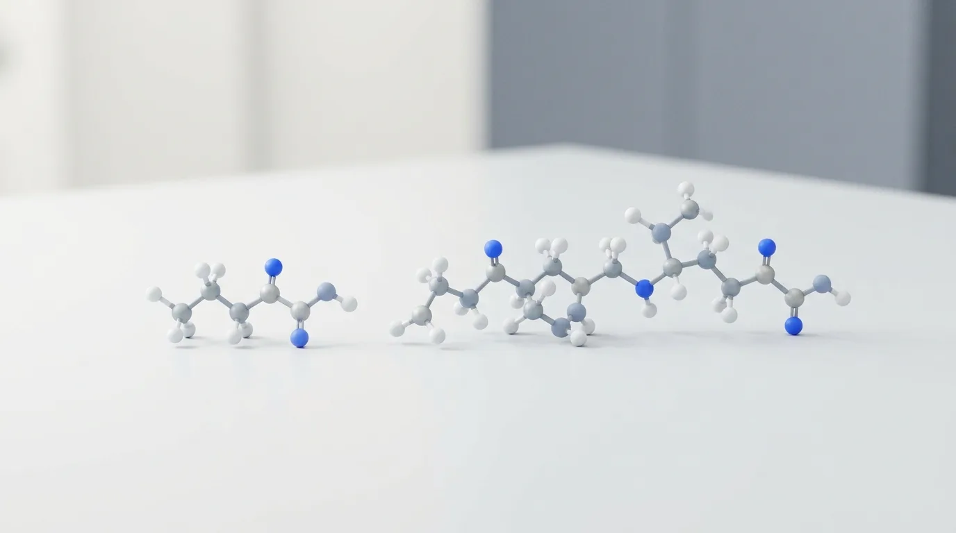
Evidence asymmetry is pronounced across the melanocortin literature. While synthetic structural analogs of alpha-MSH (such as afamelanotide, which substitutes norleucine at position 4 and D-phenylalanine at position 7) have advanced through formal human clinical development programmes for erythropoietic protoporphyria, unmodified native alpha-MSH and the truncated KPV tripeptide have zero human efficacy trials behind them. Comparing KPV against full-length alpha-MSH in the table below sets preclinical assay behaviors side by side rather than comparing equivalent clinical datasets.
| Research attribute | KPV (alpha-MSH 11-13) | Full-length alpha-MSH (1-13) |
|---|---|---|
| Amino acid residue count | 3 residues (Lys-Pro-Val) | 13 residues (acetylated tridecapeptide) |
| Primary cellular target or transporter | PepT1 (hPepT1) oligopeptide transporter | Melanocortin receptors (MC1R, MC3R, MC4R, MC5R) |
| Canonical second messenger response | Receptor-independent IkB-alpha stabilization (no cAMP surge) | Gs-protein activation elevating intracellular cAMP |
| Melanogenesis stimulation in vitro | Absent in melanocyte and MC1R-null assays | Active via MC1R agonism |
| Dominant signaling inhibition | NF-kB nuclear translocation and MAPK phosphorylation | NF-kB suppression secondary to melanocortin receptor engagement |
| Highest published trial stage | Preclinical rodent and ex-vivo tissue models only | Preclinical and ex-vivo human cell assays (unmodified peptide) |
| US regulatory status (as of Nov 2026) | Not FDA-approved; temporary 503A enforcement discretion | Not FDA-approved for clinical indications |
Researchers consulting our in-depth compound research guides frequently note that truncating 10 amino acids removes susceptibility to several endopeptidases that cleave central alpha-MSH bonds, while simultaneously creating a dependency on di- and tripeptide brush-border transporters. Where PepT1 is absent, unmodified KPV shows minimal passive membrane permeability due to its hydrophilicity.
What have rodent colitis and mucosal barrier assays measured?
Oral and nanoparticle-encapsulated KPV formulations have reduced colonic myeloperoxidase (MPO) activity, attenuated body weight loss, and preserved tight junction proteins (occludin and ZO-1) in dextran sulfate sodium (DSS) and 2,4,6-trinitrobenzenesulfonic acid (TNBS) murine colitis models. Every intestinal barrier outcome reported to date originates from cultured cell monolayers or rodent experiments.
In the foundational 2008 Gastroenterology investigation by Dalmasso et al., mice challenged with DSS or TNBS received oral KPV added to drinking water. Treated mice exhibited lower histological inflammation scores, reduced colonic MPO enzymatic activity (a direct biochemical marker of neutrophil infiltration), and suppressed mucosal mRNA levels of IL-1beta, IL-6, TNF-alpha, and IFN-gamma compared to untreated colitic controls.
Crucially, those protective effects disappeared in PepT1-knockout models or when transport was blocked, tying in-vivo tissue preservation directly to oligopeptide symport. In parallel polarized epithelial monolayer assays, KPV exposure supported the structural localization of tight junction proteins occludin and zonula occludens-1 (ZO-1), limiting paracellular flux across damaged enterocyte sheets.
In experimental murine colitis models reported in Gastroenterology (Dalmasso et al., 2008), nanomolar PepT1-mediated uptake of the KPV tripeptide suppressed colonic myeloperoxidase activity and inhibited both NF-kB and MAP kinase inflammatory signaling cascades without requiring functional MC1R expression.
Because free oral KPV undergoes rapid enzymatic hydrolysis and premature absorption in the upper small intestine before reaching the distal colon, pharmaceutical scientists have engineered targeted delivery matrices. In 2010, Laroui et al. loaded KPV into biodegradable polylactide nanoparticles collapsed inside a polysaccharide (alginate-chitosan) hydrogel to restrict payload release to the mouse colon, reporting colitis attenuation at drug concentrations orders of magnitude lower than free KPV solution. Fourteen years later, biomaterial engineering expanded on this exact delivery bottleneck:
- Polysaccharide and polymeric nanocarriers (2024): Marotti et al. (2024) evaluated a nanoparticle platform designed for combined mucosal healing and immunomodulation in preclinical inflammatory bowel models.
- PepT1-targeted co-assembled nanodrugs (2024): Zhang et al. (2024, Frontiers in Pharmacology) developed a PepT1-targeted nanodrug co-assembling an anti-inflammatory peptide with an immunosuppressant to evaluate combined mucosal repair in acute and chronic DSS-induced mouse colitis.
- Inflammation-triggered oral conjugates (2026): Cheng et al. (2026, Science Advances) engineered inflammation-triggered self-immolative conjugates designed to overcome gastrointestinal enzymatic barriers during oral peptide delivery in preclinical animal tests.
Put simply, unprotected KPV breaks down quickly in the upper digestive tract, prompting material scientists to wrap the tripeptide in protective hydrogels and smart nanoparticles so it survives transit to inflamed mouse colon tissue.
Beyond the gut: What do cutaneous, ocular, and antimicrobial models show?
Outside gastrointestinal assays, preclinical studies have tested KPV and dimeric derivatives across microporated human skin, fine-dust-stressed human keratinocytes, rabbit corneal epithelium, and fungal pathogen cultures. None of these non-intestinal investigations involve human clinical efficacy trials, and several rely on chemically modified KPV dimers rather than the monomeric tripeptide.
Cutaneous stress and transdermal permeation represent active branches of KPV laboratory study. Because KPV is hydrophilic and zwitterionic at physiological pH, passive diffusion across the intact lipophilic stratum corneum is poor. Pawar et al. (2017) investigated transdermal iontophoretic delivery and microneedle microporation across excised human skin in vitro, demonstrating that electrical current and microchannel formation substantially enhanced transdermal KPV flux compared to passive diffusion.
More recently, Sung et al. (2025) exposed cultured human keratinocytes to particulate fine dust in vitro; KPV treatment mitigated keratinocyte apoptosis and reduced reactive oxygen species accumulation by downregulating MAPK and NF-kB phosphorylation.
Ocular, vascular, and host-defense assays broaden the experimental scope while introducing important structural distinctions. In a 2006 Experimental Eye Research study, Bonfiglio et al. applied the COOH-terminal tripeptide alpha-MSH(11-13) topically to rabbit corneal epithelial defects, observing accelerated re-epithelialization that was abolished by nitric oxide synthase inhibitors, implicating nitric oxide signaling in rabbit ocular wound closure. In vascular biology, Zhang et al. (2024, Advanced Healthcare Materials) co-assembled KPV with rapamycin into carrier-free nanodrugs to study vascular smooth muscle calcification in preclinical models.
Researchers must also distinguish monomeric KPV from cysteine-bridged dimers such as (CKPV)2. Cutuli et al. (2000) reported direct in-vitro candidacidal and antimicrobial effects of alpha-MSH peptides against Candida albicans and Staphylococcus aureus, while Catania et al. (2005) solved the three-dimensional NMR solution structure of the dimeric candidacidal analog [Ac-CKPV]2, and Gatti et al. (2006) tested (CKPV)2 against endotoxin-induced host reactions in surgical rodent models. Monomeric KPV and disulfide-linked [Ac-CKPV]2 have distinct spatial conformations; antimicrobial potencies measured with the dimer cannot be automatically extrapolated to unbridged monomeric KPV.
What changed in FDA 503A policy for KPV in late 2026?
As of November 2026, KPV is not FDA-approved for any human clinical indication, though an October 28, 2026 FDA enforcement discretion policy temporarily permits state-licensed 503A compounding pharmacies to compound KPV under strict sourcing controls while formal notice-and-comment rulemaking proceeds. Regulatory permission for pharmacy compounding does not establish clinical efficacy or safety.
Regulatory oversight of KPV shifted twice during the second half of 2026. First, on July 23, 2026, the FDA Pharmacy Compounding Advisory Committee (PCAC) voted 8-6 (with 1 abstention) to recommend placing KPV free base and KPV acetate on the Section 503A Bulks List. Following that split advisory vote, the agency issued an October 28, 2026 enforcement discretion update covering select bulk drug substances under evaluation for 503A compounding.
Academic and regulatory reviewers emphasize three critical boundaries surrounding this October 2026 policy:
- Zero human clinical trial data: As documented in independent November 2026 regulatory summaries, FDA briefing materials explicitly note that zero published human clinical efficacy trials or controlled human pharmacokinetic exposure datasets exist for KPV by oral, topical, or parenteral routes.
- Pending formal rulemaking: Enforcement discretion is an interim administrative posture rather than permanent codification on the 503A Bulks List, and individual State Boards of Pharmacy retain independent statutory authority to restrict compounding within their jurisdictions.
- Anti-doping and sports scrutiny: Coutinho et al. (2026) reviewed the proliferation of unapproved experimental peptides across recreational and professional athletics, underscoring why investigational sequences remain subject to sports governance scrutiny and strict laboratory-only chemical boundaries.
For non-clinical investigators, the distinction is absolute. Bulk chemical reagents sold by a research peptide supplier are designated strictly for in-vitro assays and animal experimentation, completely separate from 503A pharmacy prescription frameworks.
How should laboratories verify KPV purity and analytical stability?
Analytical laboratories verify KPV research material through lot-specific reversed-phase high-performance liquid chromatography (HPLC) targeting a ≥99% purity standard alongside electrospray ionization mass spectrometry (ESI-MS) molecular ion confirmation. Verified stability-indicating assays are essential because aqueous KPV undergoes pH- and temperature-dependent degradation in biological homogenates.
Pawar et al. (2015) developed and validated a stability-indicating HPLC assay specifically for lysine-proline-valine in aqueous buffers and skin homogenates, demonstrating how chromatographic separation resolves intact KPV from hydrolytic and oxidative degradation products generated by tissue peptidases. Proline at position 2 introduces conformational rigidity and cis-trans isomerization behavior that can cause peak broadening or split peaks on poorly equilibrated C18 columns if mobile-phase trifluoroacetic acid (TFA) ion-pairing and column temperature are not controlled.
What makes a research peptide batch reproducible across mucosal assays?
Assay reproducibility across cell-culture replicates depends on verifying counter-ion identity (free base versus acetate or TFA salt), residual solvent limits, and chromatographic purity via an independent, lot-matched Certificate of Analysis (CoA). Residual trifluoroacetic acid above trace thresholds can acidify unbuffered cell media and induce cytotoxic artifacts in sensitive Caco-2BBE or keratinocyte monolayers, mimicking or masking NF-kB modulation.
At Peptide.Express, every lyophilized research compound undergoes rigorous purity standards and quality assurance protocols before release to institutional and private laboratories. Procurement officers can inspect third-party HPLC lab results and Certificates of Analysis matched by batch number, confirming ≥99% purity by HPLC alongside mass spectrometry identity verification from named analytical testing facilities. Supplying lyophilized peptides sealed under inert atmosphere protects the hygroscopic Lys-Pro-Val sequence from moisture-driven diketopiperazine cyclization or proteolytic contamination during cold-chain transit.
Significant knowledge gaps remain open for future preclinical exploration. Investigators have yet to isolate the exact cytosolic binding protein responsible for IkB-alpha stabilization once KPV exits the PepT1 channel, nor have comparative toxicokinetic profiles been published across extended multi-month animal exposure protocols. Addressing those mechanistic unknowns requires rigorously characterized analytical reagents, transparent negative controls (such as PepT1-deficient cell lines or scrambled VPK tripeptides), and strict adherence to laboratory-only research standards.
References
- Read the Dalmasso 2008 record on PubMed: PepT1-mediated tripeptide KPV uptake reduces intestinal inflammation
- Read the Getting 2003 record on PubMed: Dissection of the anti-inflammatory effect of the core and C-terminal (KPV) alpha-melanocyte-stimulating hormone peptides
- Read the Laroui 2010 record on PubMed: Drug-loaded nanoparticles targeted to the colon with polysaccharide hydrogel reduce colitis in a mouse model
- Read the Sung 2025 record on PubMed: Lysine-Proline-Valine peptide mitigates fine dust-induced keratinocyte apoptosis and inflammation by regulating oxidative stress and modulating the MAPK/NF-κB pathway
- Read the Cheng 2026 record on PubMed: Inflammation-triggered self-immolative conjugates enable oral peptide delivery by overcoming gastrointestinal barriers
- Read the Pawar 2017 record on PubMed: Transdermal Iontophoretic Delivery of Lysine-Proline-Valine (KPV) Peptide Across Microporated Human Skin
- Read the Pawar 2015 record on PubMed: Stability-indicating HPLC assay for lysine-proline-valine (KPV) in aqueous solutions and skin homogenates
- Read the Elliott 2004 record on PubMed: alpha-Melanocyte-stimulating hormone, MSH 11-13 KPV and adrenocorticotropic hormone signalling in human keratinocyte cells
- Read the Bonfiglio 2006 record on PubMed: Effects of the COOH-terminal tripeptide alpha-MSH(11-13) on corneal epithelial wound healing: role of nitric oxide
- Read the Kelly 2006 record on PubMed: Immobilized alpha-melanocyte stimulating hormone 10-13 (GKPV) inhibits tumor necrosis factor-alpha stimulated NF-kappaB activity
- Read the Marotti 2024 record on PubMed: A nanoparticle platform for combined mucosal healing and immunomodulation in inflammatory bowel disease treatment
- Read the Zhang 2024 record on PubMed: PepT1-targeted nanodrug based on co-assembly of anti-inflammatory peptide and immunosuppressant for combined treatment of acute and chronic DSS-induced ColitiS
- Read the Zhang 2024 record on PubMed: KPV and RAPA Self-Assembled into Carrier-Free Nanodrugs for Vascular Calcification Therapy
- Read the Cutuli 2000 record on PubMed: Antimicrobial effects of alpha-MSH peptides
- Read the Catania 2005 record on PubMed: Three-dimensional structure of the alpha-MSH-derived candidacidal peptide [Ac-CKPV]2
- Read the Gatti 2006 record on PubMed: Inhibitory effects of the peptide (CKPV)2 on endotoxin-induced host reactions
- Read the Ichiyama 2000 record on PubMed: The neuroimmunomodulatory peptide alpha-MSH
- Read the Catania 2000 record on PubMed: The neuropeptide alpha-MSH in host defense
- Read the Coutinho 2026 record on PubMed: A new era of doping? Use of peptide and peptide-analog drugs in recreational and professional sport and bodybuilding: a critical review